1/3
A. Axial imaging before and after the onset of parkinsonian symptoms in a person with MS who developed hemiparkinsonism.
B. Coronal imaging before and after the onset of parkinsonian symptoms in a person with MS who developed hemiparkinsonism

2/3
A. Axial T2 FLAIR section of brain MRI after person with MS developed right-sided hemiparkinsonism
B. Coronal T1 MRI section after person developed right-sided hemiparkinsonism. A new left hyperintense lesion is visible in the left globus pallidus with peripheral ring enhancement.
C. Axial FLAIR MRI shows a left pallidal lesion in a person with MS who developed right-sided hemiparkinsonism.
D. Nonenhancing left pallidal lesion in a person with MS who developed hemiparkinsonism.

3/3
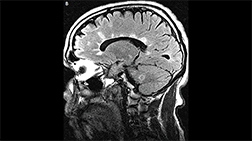
Medial longitudinal fasciculus lesion. A vertical lesion in the central midbrain involves the medial longitudinal fasciculus near the dorsal edge and spreads all the way to the ventral surface giving an appearance of a split midbrain. The right temporal lobe subarachnoid cyst is an incidental finding.