1/10
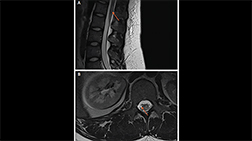
A. Longitudinally Extensive Transverse Myelitis (LETM). Sagittal T2 MRI of the cervical spine shows extensive cord signal abnormality.
B. Sagittal T1 postgadolinium MRI of the spine shows patchy enhancement.
C. Axial T2 MRI of the cervical and thoracic spine shows abnormal signal intensity located centrally in the spinal cord as is characteristic in NMOSD.
D. Sagittal T2 MRI of the thoracic spine shows extensive cord signal abnormality.
E. Sagittal T2 MRI of the thoracic spine shows extensive cord signal abnormality.

2/10
Longitudinally extensive T2 hyperintense spinal cord lesion in neuromyelitis optica spectrum disorder (NMOSD).

3/10
A. Spinal cord lesion in sagittal plane in antimyelin oligodendrocyte glycoprotein (MOG) antibody-associated disease
B. Spinal cord lesion in transverse plane in antimyelin oligodendrocyte glycoprotein (MOG) antibody-associated disease

4/10
Brain MRI from Mr. L’s second hospitalization demonstrates resolution of the previously visualized right temporal lesion and leptomeningeal enhancement (A), with an enhancing lesion in the spinal cord.

5/10
Axial fluid-attenuated inversion recovery (FLAIR) MRI of the T2 parasagittal imaging of the cervical spine from a boy, age 16 years, with multiple sclerosis. Note the high lesion burden and areas of early confluence. Children with MS are subject to more frequent relapses and typically have a higher lesion burden than adults with MS. (Images courtesy of Dr. Aaron Carlson.)

6/10
Axial fluid-attenuated inversion recovery (FLAIR) MRI of a patient with AQP4+ NMOSD and area postrema syndrome demonstrating T2 hyperintensity in the dorsal medulla (image courtesy of Dr. Divyanshu Dubey).

7/10
Cervical spine sagittal STIR MRI of a patient with AQP4+ NMOSD with longitudinally extensive myelitis.

8/10
Cervical spine sagittal STIR MRI of a patient with AQP4+ NMOSD with longitudinally extensive myelitis including an area of contrast enhancement on T1 MRI post-gadolinium.

9/10
MRI cervical and thoracic spine showing a T2 to T5 longitudinally extensive hyperintense lesion consistent with the diagnosis of neuromyelitis optica.

10/10
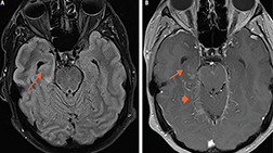
Nerve root entry zone lesion. Arrow: Lesion along left trigeminal root; the trigeminal nerves are seen in the prepontine cisterns.