1/4
A. Brain MRI from Mr. L’s initial hospitalization with increased signal intensity in the right temporal lobe
B. Brain MRI with enhancement of the temporal lesion (small arrow) with leptomeningeal enhancement (large arrow).

2/4
Brain MRI from Mr. L’s second hospitalization demonstrates resolution of the previously visualized right temporal lesion and leptomeningeal enhancement.

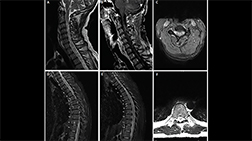
3/4
Typical lesions of multiple sclerosis are found in the infratentorial regions.

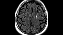
4/4
Inferior temporal lobe lesion. An inverted J lesion is in the left inferior temporal lobe, and a subtler lesion is in the right temporal lobe. Note the peripheral brainstem lesion in the left midbrain and a lesion in the left temporal cortex.