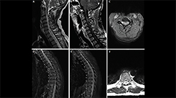
1/4
Longitudinally extensive enhancing optic nerve lesion in neuromyelitis optica spectrum disorder (NMOSD)

2/4
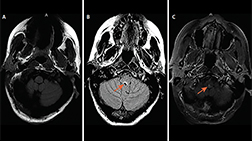
Brain MRI from Mr. L’s second hospitalization demonstrates resolution of the previously visualized right temporal lesion and leptomeningeal enhancement with new enhancing lesions both infratentorially bilateral optic neuritis.

3/4
Brain MRI from Mr. L’s second hospitalization demonstrates resolution of the previously visualized right temporal lesion and leptomeningeal enhancement with new enhancing lesions both infra- and supratentorially bilateral optic neuritis with greater enhancement in the right optic nerve compared with the left.

4/4
A. Axial T1 fat-saturated post-gadolinium MRI in a patient with AQP4+ NMOSD demonstrates bilateral optic neuritis over more than 50% of the optic nerves (arrows).
B. Optical coherence tomography taken 1 year after bilateral optic neuritis demonstrates thinning of the right and left retinal nerve fiber layers (right 62 μm, left 54 μm; normal thickness on OCT is more than 80 μm; OCT images contributed by Dr. Sara Qureshi).